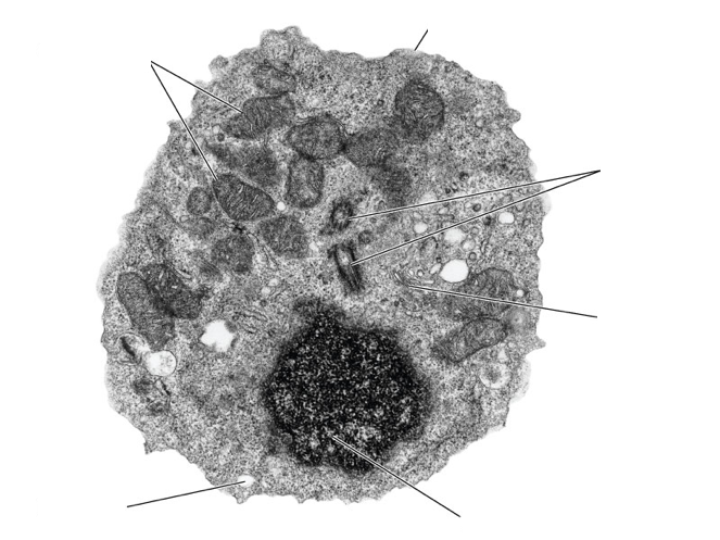
animal cell under electron microscope
How would you identify if a cell was a plant or animal cell when looking at it under a microscope. Light and electron microscopes allow us to see inside cells.
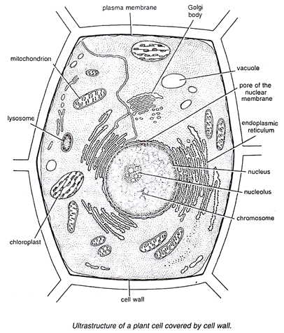
The Cell Form 1 Biology Notes Easy Elimu
Animal Cell Under An Electron Microscope.
. An animal cell also contains a cell membrane to keep all the organelles. Viewing Animal Cells under a microscope. Plant animal and bacterial cells have smaller components each with a specific function.
Because of this dimension they are not visible under optical microscope. Animal cells have a basic structure. A typical animal cell as seen in an electron microscope Medical Images For PowerPoint 1.
Electron Microscopic Study Of Cell And Organelles Important The largest known animal cell is the ostrich egg which can stretch. Beneath a plant cells cell wall is a cell membrane. Most animal cells are between 001 mm 005 mm.
Structure of plant and animal cells under an electron microscope Advanced Higher Biology Cell and molecular Biology The Electron Microscope Two main advantages High resolving power. Below the basic structure is shown in the same animal cell on the left viewed with the light microscope and on the right with the transmission electron. Animal cell under electron microscope.
Animal cell under electron. Typical Animal Cell Pinocytotic vesicle Lysosome Golgi vesicles Golgi vesicles. Observing a wide range of biological processes and animal cell under light microscope is easier due to advances in microscopic techniques.
Beneath a plant cells cell wall is a cell membrane. However their presence was inferred long before their observation due to plasmolysis bursting of animal cells placed. An animal cell also contains a cell membrane to keep all the organelles and cytoplasm contained but it lacks a cell wall.
Under a microscope plant cells from the same source will have a uniform size. Beranda Animal Cell Structure Under Electron Microscope Difference Between Plant And Animal Cells Cells As The Basic Units Of Life Siyavula Because of the limited. A typical animal cell as seen in an electron microscope Medical Images For PowerPoint 1.
Electron microscopes are used to investigate the ultrastructure of a wide range of biological and inorganic specimens including microorganisms cells large molecules biopsy.
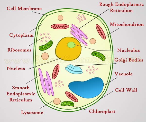
Structure Of Animal Cell And Plant Cell Under Microscope Diagrams

13 Draw A Well Labelled Diagram Of A Generalized Typicalanimal Cell As Seen Under Electron Brainly In

Anatomy And Physiology Of Animals The Cell Wikieducator
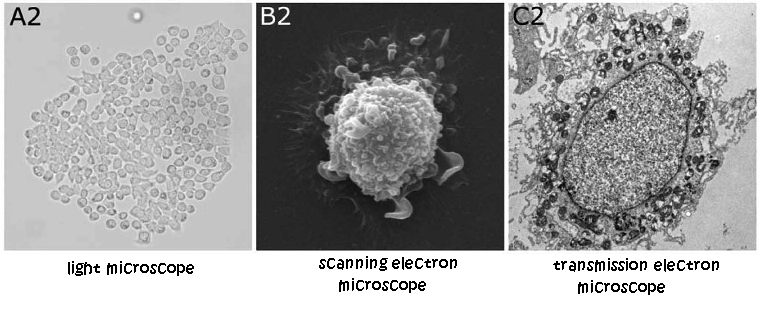
Topic 1 2 Ultra Structure Of Cells Amazing World Of Science With Mr Green
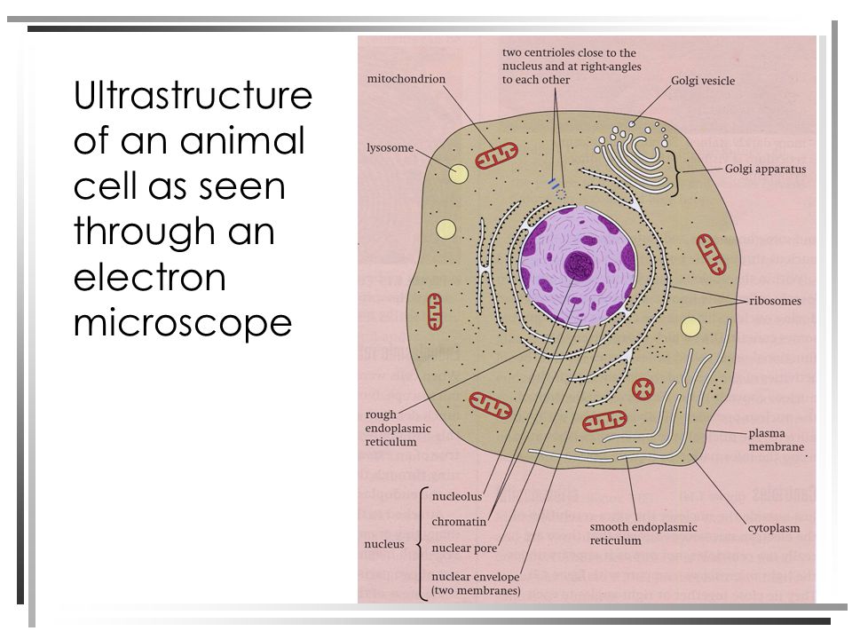
Structure Of Plant And Animal Cells Under An Electron Microscope Ppt Video Online Download

Q14 Draw A Large Diagram Of An Animal Cell As Seen Through An Electron Microscope Label The Parts Brainly In

A Typical Animal Cell As Seen In An Electron Microscope Medical Ima
K C S E Biology Q A Model 2013pp1qn02 Atika School

Illustrate Only A Plant Cell As Seen Under Electron Microscope How Is It Different From Animal Cell

Scanning Electron Microscopy Of Cells Grown In Vitro And Of Giant Download Scientific Diagram
Cil 7724 Rattus Epithelial Cell Epithelial Cell Of Thymus Cil Dataset
Free Ncert Solutions For 9th Class Science The Fundamental Unit Of Life Studyadda Com



Comments
Post a Comment